Diaphragm Of A Microscope
What is the diaphragm of a microscope, and how does it work? You’d have to say two things must perform exceptionally well if a microscope is going to function as it should.
First of all, light must hit the object being viewed, and secondly, once the light has illuminated the specimen, it must be collected and magnified.
One component essential to this procedure is the iris diaphragm.
In this article, we’ll discuss what exactly the iris diaphragm is, how it works, and its responsibilities.

What Is A Diaphragm?
A diaphragm on a microscope is responsible for how much light leaves the microscope condenser. This light controls the illumination of the object in view. In addition to that, it simultaneously controls the contrast between the background and the specimen.
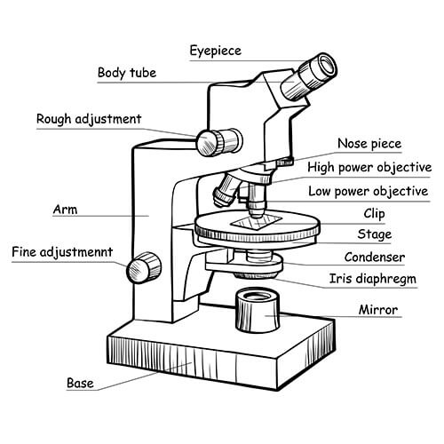
To make this work, in the center of the diaphragm is a round hole called an aperture. The iris diaphragm controls the aperture size, which is where the light passes through. The diaphragm sits between the condenser and the specimen.
The smaller the aperture on the iris diaphragm, the less light can pass through. In effect, this reduces the illumination of the specimen but increases the contrast.
Moreover, there’s a direct comparison between the expansion and contraction of the microscope’s diaphragm, which is similar to that of the eye’s iris.
What Is An Iris Diaphragm?
The iris diaphragm is a design that allows adjustment and size of the aperture opening; as we previously mentioned, the name iris is taken from the eye.
There are several types of iris diaphragms; the most popular and widely used ones utilize a set of blades in a circular arrangement. Controlling the blades affects the size of the aperture.
Another well-known diaphragm is a Zeiss diaphragm that rotates. This diaphragm type has a circular disc in which are several apertures, all varying in size. Another type of diaphragm is the Waterhouse. This employs a set of diaphragms made from brass strips that you can use interchangeably.
What Is The Purpose Of The Iris Diaphragm?
Microscope Illumination
The primary responsibility of the iris diaphragm is controlling how much light hits the specimen. The light in question is the microscope’s primary light source, and this is collected by the microscope’s condenser, subsequently regulated by the diaphragm before shining through the specimen.
Opening the aperture in the iris diaphragm wider will intensify the amount of illumination reaching the specimen; Simultaneously, this makes the image brighter. The light is not so focused, and that reduces the contrast.
Ideally, you need the iris diaphragm open sufficiently wide enough to illuminate the specimen. Hence, the image is bright, but you don’t want to open the diaphragm to such an extent the sample looks washed out and anemic looking.
Microscope Focus
To some extent, the iris diaphragm can influence the specimen’s image focus. The amount of contrast between the background and sample and within the confines of the sample itself is an essential factor.
Most microscope iris diaphragms contain between five and ten blades; however, diaphragms can be as few as two and upwards to twenty blades. Because the diaphragm is a circle curving the blades make a more circular opening, whereas straight blades generate a multifaceted shape.
Increasing the number of blades in the iris diaphragm means the opening is much more circular resulting in higher contrast and more focused light. Perhaps unsurprisingly, these iris diaphragms are more expensive to make and therefore are typically found on more elaborate and advanced equipment.
How Does An Iris Diaphragm Work?
As we previously discussed, the iris diaphragm controls illumination and contrast levels on the magnified image of an object being viewed. Open the diaphragm’s aperture wider if you want more illumination and lower contrast, and so forth.
Diaphragms are positioned low down near the bottom of microscopes. They need to be above the light source and condenser but below the specimen stage. Iris diaphragms are opened by turning them clockwise to close and counterclockwise when you wish to open them.
It’s also possible to find iris diaphragms inside the condenser; if your microscope has this configuration, it’s known as an Abbe condenser. Moreover, the diaphragm control will be on the condenser.
Brightness Vs. Iris Diaphragm
Most importantly, you should remember the iris diaphragm isn’t responsible for the light’s intensity. This is the responsibility of the actual light source and how it’s set on the condenser.
Iris diaphragms are limited to establishing how wide the light beam passes through the specimen, and this can only determine the amount of illuminated sample.
Magnification Vs. Iris Diaphragm
Managing the contrast by controlling how much the diaphragm illuminates the specimen is crucial in specimens’ high and intermediate magnification. Basically, when using higher magnification levels, less light can pass through; therefore, iris diaphragms must have wider openings to allow more light through.
Types of Diaphragms
Disc Diaphragm
Disc diaphragms are not as popular as the iris type and also less sophisticated in design. By and large, disc diaphragms are simply spinning wheels containing different diameter openings. Should you require more light, move the disc to a larger diameter. For less light, go smaller.
Field Diaphragm
These types of diaphragms you will find closer to the light source of the microscope. Field diaphragms work similarly, except they control the amount of light and the field of view size for the image. However, the diaphragm does affect the amount of light entering a microscope but not light quality or the contrast.
Conclusion
There are always compromises when using a microscope; if you’re trying to obtain the best possible and clear image, you will need to find a good balance between contrast and final appearance. It will not be possible to open a diaphragm fully and at the same time get high contrast.
As we discussed, the wider the diaphragm, the more illumination you put on the specimen, the less contrast you’ll get, and so forth. Focusing microscopes can take a long time because you’re always attempting to find the ideal balance between contrast, brightness, and the image size you can obtain.